A cerebrospinal fluid (CSF)-venous fistula is a specialized cause of spontaneous intracranial hypotension. It can be challenging to find because CSF passes directly into a vein rather than collecting in a way that routine spine imaging readily shows. Diagnosis and treatment therefore often involve a team experienced with CSF-leak imaging, neurology, neurosurgery, and endovascular care.
What a CSF-venous fistula is
CSF normally surrounds and cushions the brain and spinal cord. A CSF-venous fistula is an abnormal connection between the CSF-containing space around the spinal cord and an adjacent vein. Through that connection, CSF can move into the venous system and contribute to symptoms associated with low CSF volume or pressure.
Headache that changes with position is a common reason for evaluation, but symptoms vary and can overlap with other conditions. A fistula cannot be diagnosed from a symptom pattern alone.
Why diagnosis can be difficult
Unlike some spinal CSF leaks, a CSF-venous fistula may not create a visible collection of fluid outside the normal CSF space. Brain or spine MRI may raise suspicion for spontaneous intracranial hypotension without showing the precise connection. Symptoms and imaging findings can also vary over time.
The evaluation begins by considering the full clinical history and available brain and spine imaging. Other causes of similar symptoms may need to be assessed. When a fistula remains a concern, specialized imaging can be planned to look for the brief passage of contrast from the CSF space into a vein.
Specialized imaging
Dynamic CT myelography and digital subtraction myelography are among the techniques specialists may use to localize a CSF-venous fistula. These studies introduce contrast into the spinal CSF space and acquire images with careful timing. Positioning can help place the suspected side of the spine where the contrast is most informative.
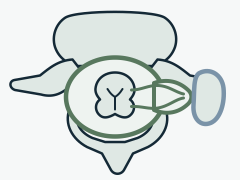
CSF stays within its normal space surrounding the spinal cord.
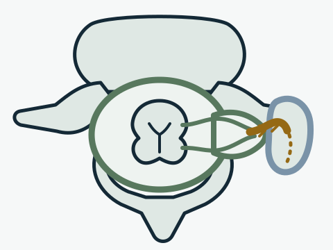
An abnormal connection lets CSF drain directly into an adjacent vein, so no fluid collects where routine imaging would show it.
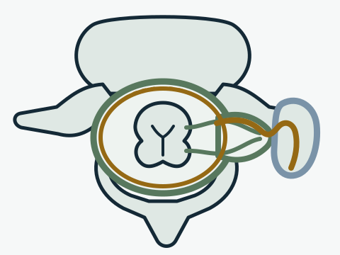
Contrast placed in the CSF space briefly outlines the abnormal connection as it passes into the vein — timing and positioning are what make the study work.
The choice of study, position, and imaging sequence depends on earlier findings and local expertise. Sometimes more than one study is needed, and a nondiagnostic examination does not by itself establish or exclude every possible cause of symptoms. The imaging team can explain the purpose and risks of the proposed procedure.
Treatment options
Treatment is individualized to the certainty and location of the diagnosis, the fistula’s anatomy, prior care, and the patient’s symptoms and health. Options discussed in specialist practice may include an epidural blood patch or targeted sealant procedure, surgical closure, and transvenous embolization. Not every option is technically available or appropriate for every fistula.
The evidence base is evolving. Much of the published experience comes from specialized centers and observational series, so treatment decisions should account for both what is known and what remains uncertain.
Surgery and transvenous embolization
Surgery approaches the involved spinal nerve-root region directly so the abnormal connection can be interrupted. The exact operation depends on the location and local anatomy. The discussion should include how the fistula was localized, what structures are nearby, the expected recovery, and procedure-specific risks.
Transvenous embolization reaches the draining vein through the venous system and places embolic material to close the abnormal pathway. Whether that route is feasible depends on the venous anatomy and the confidence of fistula localization. Surgery and embolization have different access routes, risks, and follow-up considerations; neither is a universal choice.
Recovery and follow-up
Recovery varies with the procedure, duration and pattern of symptoms, and other health factors. Clinicians may monitor headache characteristics, neurologic symptoms, function, and selected imaging findings. A new or different headache after treatment should be discussed because pressure-related symptoms and procedure-related concerns may require reassessment.
Follow-up also provides a chance to review whether symptoms are changing as expected and whether further imaging is useful. Persistent symptoms do not identify the cause by themselves, and decisions about additional testing or treatment require a fresh specialist evaluation.
Questions for a specialist
CSF-venous fistula evaluation and embolization are part of Dr. Oushy’s endovascular practice.
Terms used in this article
- cerebrospinal fluid
- The clear fluid that surrounds and cushions the brain and spinal cord, often abbreviated CSF.
- embolization
- A procedure that closes off an abnormal blood-flow pathway from inside the vessel, using materials delivered through a catheter.
- intracranial hypotension
- A condition of low cerebrospinal fluid volume or pressure around the brain, often causing a headache that changes with position.
- myelography
- An imaging study in which contrast is placed into the spinal fluid so the fluid space and any abnormal fluid movement can be seen.